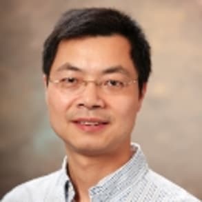
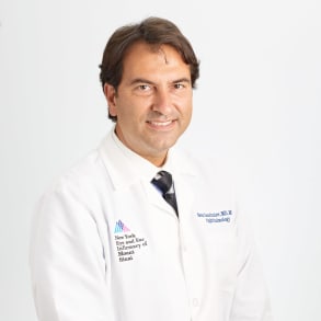
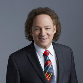
From microsurgical robotics, capable of delivering cellular therapies, to artificial intelligence and the latest imaging technology, which is mapping and identifying different patterns of visual loss and cellular changes, NYEE clinicians and scientists are pioneering the next generation of diagnostic tools and therapies, capable of treating blinding diseases such as glaucoma, diabetes, macular generation, and retinitis pigmentosa. With so many groundbreaking projects underway and the largest residency program in the nation, NYEE is the premier site for patient care, research, and training the ophthalmic leaders of tomorrow.
Refer an Ophthalmology Patient
Good afternoon and welcome to today's webinar. My name is Jim Sigh and I'm the president of new york. I your infirmary of Mount Sinai. We thank you for taking the time to learn about the past, present and future of new york i in year And we have assembled an ophthalmic dream team to share with you some of the latest breakthroughs in ophthalmology that May one day provide us with the ability to not only treat blindness and vision loss but to restore the gift of sight This year, New York I near proudly celebrates its 200 year anniversary Since its founding in 1820 as the first specialty hospital in the United States and remains one of the top ophthalmology hospitals in the nation, Ranked 11th this year by US. News World Report. Originally established as a charitable institution for the service of new york cities, core for treating diseases of the eyes, ears, nose and throat. The infirmary has become a leading clinical research and teaching enterprise. By 1918, New York India has already treated over one million patients and established the school of ophthalmology and otolaryngology. Today, new york kinda remains at the forefront of its specialties in America. New york are near, is also home to break through clinical research, which has introduced now widely practiced diagnostic and surgical techniques with a rich heritage and a mission of providing the highest quality patient care, graduate and continuing medical education, scientific research and community outreach. New york are near, has built upon its strength to maintain a leadership position in the fields of ophthalmology, on allowing ology, head and neck serve a story and plastic and reconstructive surgery. Lastly, new york kind year has been an international leader in restoring the gifts of sight and sound for people of all ages from infants to senior citizens. As we look forward to our third century of specialty care, we are evaluating how we will need to serve our communities in the future and push forward and innovate with innovative research and exceptional patient care that will set new standards for specialty hospitals. Today's webinar will provide you with a snapshot of a few of our transformational clinical and research projects which will revolutionise the practice of ophthalmology around the world. It's my pleasure to turn the webinar over to one of our co moderators. Dr Sandy Maser, professor of ophthalmology and pharmacological sciences and Vice chair for academic development and mentoring in the department of ophthalmology at the Icahn School of Medicine at Mount Sinai Dr Maser. Thank you dr CY we have a superb panel of experts lined up today. We're going to ask each of them to introduce themselves and to talk a little bit about their work here at the new york I Anier, our first Panelist is dr bo Chen. Dr Chen is an associate professor in the department of ophthalmology but also in the departments of cell developmental and regenerative biology and the nash family department of neuroscience at the Icahn School of Medicine. Dr Chen please. Yeah, thank you Doctor Blazer for the introduction and it's a great pleasure to present our Euro region generation research to come back in the house. Retinal degenerative diseases. Look at general diseases, often age related chronic conditions. The progression of of the disease takes many years, even decades take the stages of A. M. D. Age related macular degeneration. For example Patients May one Experience early signs of agent loss touching is a little Beijing and the division gets worse over time and that they are the best stage. There can be accompanied a loss of central vision. The loss of the Beijing is a result of loss of regular sales over time. Retina has shown in the bottom uh Grant panel is a piece of new york tissue in the band of I that's where we dream starts. If we take a thin slice of the retina and magnify it under a microscope. And you can see there are several types of retinal cells and the altar retina. There are photos receptor, sales, rods and cones in the inner retina. They're gathering sales, their output new round of the retina. The degeneration of the regional sales and the outer retinal photoreceptors. Sales underlying the pathogenesis of age related macular degeneration. Glaucoma is another type of leading cause of vision loss. Is the result of the degeneration of regional gathering sales. If you can think about it, the law, if the loss of the regional sales can be replenished by generating new regional sales, the vision can be restored. The question is, is that even possible? The answer is yes. Thanks to the existence of your grill sales in the reckoning. Music video sales are support cells in the retina. They are sitting in the center of the recognition. Music video sales are poor of retinal stem cells in in the presence of environmental insults or genetic defects. The loss of regional sales can be seen spend your grill sales and they occupied and making a lot of daughter sales. And those daughter sales can differentiate into regular sales to replenish lost weight in your neurons and restore vision. And this is a powerful self repair system and that that is full regeneration capability in lower species such as zebra fish. However, in the main menu retina, including labor, animals such as mice all the way up to humans. We lost the regenerative capability in europe real sales. My lab research effort is focused on unlocking the regenerative capability of urethral cells. In the main menu written to restore vision showing in the bottom left is our working mice. So we showed him for the first time that we can use gene therapy strategies to unlock and restore vision in mice. After two sequential gene transfer in builder grill sales, we can activate the music, video sales, demonstrating and generate new photo receptor cells as you can see in this red red sails here, the shape like tempo and these are new photo receptor cells and we can even restore congenital blindness in mice, our research provided first print a proof of principle. They had the reactivation of the regeneration of the Romanian retina to restore vision is possible and feasible. The ultimate goal of our research is to turn our left discoveries in mice to eventually for therapies in humans. There are a lot of steps between and the next big milestone might have been the long human primates monkeys this year. Similar feature features treats with humans and our research is one step of at the time from the research for mice and all the way up to you humans with that. I end my talk. Thanks for your attention. Thank you very much. Doctor Chen. This is your at a very exciting stage for this research. Our next Panelist is Doctor Toko too dr to is the director of the David di Maria's adaptive optics retinal imaging laboratory at the new york. I Anier and also associate professor in the department of ophthalmology at the Icahn School of Medicine. After choi um Thank you Dr Maser. Hi this is Tokyo choice. Um How's everyone doing today? Um My research at New Anya. It's about non invasive imaging of human eyes with different retinal diseases. And you may ask what is threatening imaging. So Randall imaging takes digital pictures of the back of your eyes. And these pictures helped your doctor to find certain diseases and check the condition of your eyes. And these pictures can be processed to give a lot of useful information about the health of the neural tissue and blood vessels on the back of the ice. And your doctor may recommend certain right no imaging procedures if you have any systemic conditions or eye diseases such as diabetes, uh macular degeneration or glaucoma. So in our lab we take retinal imaging into the next level by imaging the eye at a cellular level. And our research. And your idea involves two different high resolution retinal imaging devices. So the first one the first imaging device it's called adaptive optics of pharma scope. So to simply put it's a high resolution optical camera. So comparing the traditional imaging devices and this high resolution ocular camera, it's like comparing the old fashioned tip TVs to the four K ultra high definition TVs. So here on the PowerPoint slides here, I present you a case from two years ago a patient came to our clinic due to a small change of his fishing and none of the regular retinal imaging devices was able to identify what for the problem uh in the retina. And this patient was then referred to us for high resolution seller imaging. And it turned out that he had macular toxicity due to liquid Viagra overdose. And has written a picture. It's on the right panel on the proper side and which you can see a lot of dark regions um indicating individual light sensitive cells dropped out as compared to the one on the left panel which is from a healthy control. So using this cellular imaging the technique, we are able to help both the patient and the doctors to identify the problem. Um in the right now um the second imaging device is uh it's called optical coherence tomography. And again to simply put, it's like Oculus ct scan. And the technique allows us to image a very specific type of aquila in themselves on the right now. And on the left here it's uh one millimeter cube volume on the back of the eye image. Using this Oculus city scan device. And on the right panel it's an image showing the surface of this cube which is the surface of the right now. And each tiny little white dots on this image represents a single female cells. And as you can see in this movie we are able to capture the movement of these cells and these self movement and function. Really reminds me of those I robot cleaners that I have a very short movie in here. Um so they both move around on the surface and try to keep the surface cleaned. And this is how I explain to my patients about the functions of these emails cells. And we are currently applying this uh imaging approach on patients with diabetic retinopathy and glaucoma to see if we're able to use this type of email cells as a biomarker for early diagnosis of the disease is um so that's it for my part today and I will turn it over back to Dr Maser. Thank you for listening. Thank you. That was wonderful doctor too. Um our 3rd Panelist is Dr Sean Yeah Nikolaev and Dr Yang Talev is a professor in the department of Ophthalmology at the Icahn School of Medicine and is also director of the Ophthalmic Innovation and Technology Program at new york. I Anier, Dr Yan Shalev, Thank you Dr Maser and so great to be here with my esteemed colleagues and talk about research and innovation. I occupy myself more with the innovation because what is research without the innovation? If we can get it to patients, It's kind of like a car without transmission. And I'm here today to really share with you about the milestone that we reached earlier this year. And it's an interesting and very timely milestone. It comes on the 200 bicentennial for a new york Pioneer, but more importantly, it's a milestone for our department. Um it's a milestone for ophthalmology in the new york area and it happens to be also a milestone for the United States and uh there have been a lot of first through the history of ophthalmology at new york Pioneer and uh the new york area within introduction, for example of fake falsification technology, which is how cataract surgery is done. Uh And we have now the number one surgical procedure in the in the world in terms of volume is done in a technology that came out of the new york area. Uh And more recently we introduced them and installed at the new york Pioneer, the first robotic surgical system with clinical uh surgical capabilities based on a collaboration that has been In the works for the past 6, 7 years. Um And uh and this is quite pivotal I think because uh robotic surgery is really coming upon us and this is the first surgical system for atomic that has the level of precision that makes it relevant to the field of ophthalmology. So again I would also like to highlight that would not be possible without the leadership of our ophthalmology department and doctors I as well as really significant input by the Mount Sinai Innovation Partners and so many cross functional groups to really bring this cutting edge technology all the way from europe that is one of a kind in our country and the world. And I want to I will shut up for a second to play this video now because I think it really encapsulates uh that system. It's much better as a visual than as an audio. So please play the video. Yeah. Mhm. Yeah. Mm. Yeah. Mhm. And. Mhm. Yeah mm mm. Yeah. Yeah. Yeah mm. Yeah. Mhm. Thank you. So as you see the robotic installation is quite different in the way we do atomic surgery. And it happens to be also a platform technology. And what does platform technology mean? It means that it goes beyond a single application in this case. This can be impactful for everything from retina surgery where we need high precision uh intervention to cataract surgery, to glaucoma to cornea. It can really touch every aspect of surgical intervention. And very often I'm asked well, how can why is robotic surgery necessary? And uh and in terms of the scale and the impact? Probably the best way to illustrate that is if you think about surgical precision of a very good surgeon uh in ophthalmology, that precision is about 100 microbes. The resolution of that precision is about 100 microns of how fine of a discrimination one can have. That surgical system takes us down To about 5-10 microns almost 10 times better. And when it comes to ophthalmology we know smaller is better and more precise is better as well. So we're very excited this system is going to go into FADA clinical trials. It's already been approved in Europe. Um and we're trying to initiate the FDA clinical studies here in the US so that we can lead the way of multiple applications of this high precision micro intervention in the town field. And that would be a collaborative effort with a lot of the faculty here at new york pioneering Mount Sinai. And again, this will be something that really in my mind illustrates the resilience and the innovation that we have that in the middle of a pandemic, in the middle of a pandemic from ground zero margin april were tough months in the new york, as we all know, we were able to bring that system in and install it virtually because people couldn't travel from the Netherlands and really get it operational here. So it's very exciting And this is one aspect of all the innovation that we're doing at the cutting edge and really driving the next wave for the next centennial. Thank you very much. Thank you daddy. And so this has been very exciting so far and I now want to turn over the program to dr paul Saudati, my co moderator, to introduce the remaining panelists. Thank you Dr mazer and good afternoon everyone. Our next Panelist is Dr Richard Rosen. Dr Rosen is the pierce. Distinguished Chair in ophthalmology. Deputy Chair for Clinical Affairs. Vice chair for ophthalmology research surgeon, Director and rental service chief at new york. I. Anier and Professor in the department of Ophthalmology at the Icahn School of Medicine. Thank you. Dr Sidoti. Great. I'm gonna talk about quantitative assessment of the retinal blood vessels. The i is the one place where we can directly visualize the central nervous system and its capillaries by looking through the pupil were able to see the optic nerve which is part of the brain. And we can see all of the blood vessels with optical coherence. Tomography angiography, a new non invasive technique. We're able to visualize the retinal vessels without the use of any external um contrast agent. We can break down the blood vessels into multiple layers which is something we've never been able to do before. And it allows us to distinguish changes between a normal and a diabetic. I so here you can see in a patient with diabetic retinopathy there are small micro aneurysms, there's loss of capillaries. And when you compare this to a normal you can see that the diabetic eye has lost. It's somewhat threadbare and it's um amount of capillaries, their small little bulbs there which you can appreciate as being abnormal blood vessels that have closed off compared to this healthy i what you're seeing here on the right. So as you go through the different layers you can appreciate how the disease has really ravaged through the the I we've in our lab we've actually developed a number of quantitative measures looking at the density of blood vessels, looking at the blood oxygen supply. Changes in the structure, loss of blood vessels uh changes in the overall appearance and in the risk for progression. We've actually added this to the clinical measures to try to see how diabetic retinopathy and glaucoma progress in a vascular manner. So the, okay so this we've added adaptive optics and geography that dr chui showed you. And basically with adaptive with O. C. T. Angiography we capture individual frames of a movie. So sometimes certain vessels will appear and sometimes will disappear. And we've used this to study sickle cell disease. Sickle cell disease. Most common inherited blood disease and basically involves abnormally shaped blood vessels which include the small capillaries. You can actually see that there is intermittent and flow in this central area within the retina. And you can with the adaptive optics were actually able to see individual red blood cells as they track through these capillaries and see that the changes that occur. This is actually a single abnormally shaped cell in capillary. In patients. I we've taken this one step further to try to develop a means of measurement of therapeutic value of certain things. And here's a patient who has been treated pre treatment and post treatment. And you can see that with treatment we've actually they've actually been able to decrease the intermittent flow in this patients. I when we looked at the closer with adaptive optics were actually able to appreciate what kinds of changes occur. So we have areas where the blood is blocked and sometimes it's restored. Other areas where the capital is completely closed off and it's restored. In other areas. We look and we see that their pre and post treatment there may be restoration. There may also be um areas that are regress and in fact this treatment is not perfect. So in some cases the disease still continues. So in summary uh the high resolution imaging that we've been involved in developing shows us many new aspects of disease that clinicians really were not able to access in patients and will help lead us into the next generation of therapy for some of these very important diseases. Thank you for your attention, That's really fascinating work. Dr Rosen, thank you. R 5th panelists today is Dr Luis Pascual. Dr squali is a professor in the department of ophthalmology at the Icahn School of Medicine site share for ophthalmology at Mount Sinai Hospital, Director of the I envision Research institute at Mount Sinai in new york. I near deputy chair for Ophthalmology research for the Mount Sinai health System. Thank you Dr Sidoti. People often ask um what is artificial intelligence and we know that humans learn from experience, computers can learn from prior data. In fact, other researchers have shown that you can train a computer to diagnose glaucoma from a simple optical photograph. I can do it as good as or better than glaucoma expert. I have a different vision for how artificial intelligence can help and that is to understand the upstream factors that contribute to the development of primary open angle glaucoma. Primary open angle glaucoma is a disease of the optic nerve and the optic nerve is like a telephone cable that connects the eye to the brain. And in glaucoma there's a premature deterioration of the fibers in the cable. It is projected by the year 2040. That 114 million people globally will have this disease. So what do I mean by pre clinical phase, how are we going to find these upstream factors? Well, we can take any disease and put it out in a timeline for glaucoma. We presume you start out with a normal, healthy optic nerve. Then something happens and we don't know what that something is. But the first clinical signs of the disease, the development of structural damage to the optic nerve. And as the disease progresses, there is vision loss that can ultimately lead uh, to blindness. So what we're trying to do is to find out what's going on during that pre clinical phase of the disease. And what we've been doing is following large cohorts of health professionals over decades collecting data about when they developed glaucoma and along the way we asked them of what they've been eating, what they've been drinking, how much exercise they've been doing, what other medications they're taking. Also very early on. Um we did everything from take toenails samples to DNA specimens from these folks. And what I can say very briefly is, is what I've learned is that primary, open angle glaucoma is not only secondary to one thing, it's secondary, too many things and it's a disease with a strong genetic component. And this is what we're trying to use artificial intelligence to tease out what is this complex web of path of physiology around this disease. Um in glaucoma, there are many different patterns of damage that the optic nerve can undergo. You can look up at the top left and and see an optic nerve damaged with a hemorrhage on its nerve head and associated with this as early paris central visual field loss. What we're finding is is that the patterns that of damage that developing glaucoma are secondary to many mechanisms, including vascular dis regulation. And you saw the beautiful uh slides of the art ability to uh image vascular pathology. Uh And I'm very excited by that work by my colleagues, but there are other mechanisms also estrogen deficiency, mitochondrial dysfunction and oxidative stress. And so what we know is is that the different patterns of damage to the optic nerve underlie different patterns of vision loss. And what my colleagues have developed as an artificial intelligence algorithm to objectively have identify the different patterns of visual field loss that occur in glaucoma. So we've collected over 2000 cases of new onset primary open angle glaucoma. And we asked an artificial intelligence algorithm to please learn the different patterns of visual field loss that our patients are undergoing. And you see the different patterns ranging from 81 which stands for archetype one, which is normal All the way through 16 different patterns of Lost's. Thank you. And then so what can what this artificial intelligence algorithm can do is to take a visual field such as this one here where it says TD probability, where there is some damage. And basically the algorithm is recognizing there's three different patterns of loss in this patient. There is a normal pattern, There is a pattern called 85 where there's no there's damage near the nose. And then there's another pattern called 8016 where this sweeping damage across the inferior visual field. So what are some of the insights that we've gotten so far from your taking this approach? And this is basic stuff, Number one, we learned that paris central loss is common in this disease. And so that's 80 14. Another words, there's a good percentage of glaucoma patients who present with damage right in the center of their vision akin to macular degeneration. You can read any text book on glaucoma and they don't say that this happens. Uh they say that glaucoma is a disease that affects the peripheral vision and then it gangs up on the central vision at the end. But it turns out a subset of patients will get damaged early near the center of their vision. Number two, The pattern of loss that happens in one eye tends to happen in the other eye. So unfortunately, if a patient has a strong component of archetype 14 1814 in one eye and the other eye is normal, our data is telling us that it's likely if the other eye gets damaged, it's also going to get damaged in the center of the vision. Um and that is concerning and that's something we have to stop Number three is um we have African American patients in our cohorts. And what is really dramatic is that we find that they're most predisposed to the following patterns. And if you can point to these 86 and 86 Yeah, that's a bad pattern. Um that means that you've lost a lot of visual field early on your disease 80 13 And 88. So you're either losing half of the upper part of the field, half the lower field or half of the entire field. This suggests that there's a different biology operative in patients of african descent. We have to identify these patients early and we need to get on our high horse and figure out what's going on with these patients. So where do we go from here? Well, first we start to look at what we want to do is find out what are the determinants of each of the different visual field subtypes in glaucoma. We're in a pretty good position to do that because we collected blood on our participants long before they developed any of these diseases. We took that blood. We isolated the DNA. We started away to a time which is the present where we could take a very small sample of that DNA. Not even use up their whole sample and genotype them at tens of millions of locations throughout the genome to find out what are the genetic determinants for each of these subtypes. We also took the serum component from their blood and banked it away. And for a day where we could analyze thousands of metabolites all at once from a very small sample of their specimen. This is called metabolism mix. And we're hoping by using genomics and metabolism mix that we're going to be able to uh figure out what's causing each of these different subtypes of the disease. Also, we're hopeful that because these specimens were collected during the pre clinical phase of the disease will get some insights on how to prevent glaucoma from happening in the first place. Thank you for your attention. That's really great work. Thank you. Dr pasquale. And our final Panelist is Dr Douglass Frederick, Dr Frederick, assistant chief of pediatric ophthalmology and strabismus for the Mount Sinai Health System and professor of ophthalmology pediatrics and medical education at the Icahn School of Medicine. Thank you dr Sidoti and thanks to each of you for tuning in today and participating in this webinar, I'd like to talk a little bit about the third realm within which new york pioneer truly excels. We've already heard that new york Pioneer really is the best place to receive care whether you come from new york city anywhere, United States or internationally. We've also learned that new york i new york cells and research, which you've heard from the previous talks. But for 200 years, New York I near has also been the best place to obtain education to learn how to become an ophthalmologist. I want to spend a minute talking about what that path entails. So each year there are about two million young men and women who obtained their bachelor's degree at universities and colleges around the country. Of those 2,017,000 choose to pursue a career in medical in to become a position. Of those 17,000 medical school graduates. Each year, only 650 are wise enough and smart enough to choose a career in ophthalmology. Every training program looks at those 650 applicants to determine the best for their program, who would take advantage of what our training program has to offer. About 650 applicants. We choose just 10, 10 residents who will spend four years. We're learning how to become the best clinicians, the best researchers, the best edges And uh up those 10 residents. Year after their four years of trade, they will join the 24,000 ophthalmologists who are currently practicing in these states. And in each step of the way, whether it's undergraduate medical education, graduate medication or continuing medical education. New york pioneer has really been a premiere of um this education, this opportunity to continue our personal journeys in professional development so that we can become the physicians who you want to care for your eyes, who you want to come to take care of your loved ones. I want to talk about how uh we participate in the education that every part of this this path As a medical student medical spend anywhere between four and 6 years learning to become physicians. But from the very first year have faculty members who are working with these 150 Icahn School of Medicine, a Mount Sinai medical students each week from 4 to 6 hours a day, teaching them how to become not good physicians, but also to show them the opportunities that exist in ophthalmology. The east harmon health outreach project is the opportunity for medical students to work with us and faculty members in the department ophthalmology to provide care for a population here, um the Upper East Side and East Harlem patients who wouldn't get care otherwise, who can get counted by medical students, supervised by members of the faculty as well as trainees within the department of Biology. After medical school, We spent our focus training residents, residents spent four years learning to become ophthalmologists. Uh they take care of about 15,000 patients during that four years time period. Uh they received oh 500 hours of didactic education. They perform hundreds of surgeries and 1000 features during that time. That it's an intensive time period. But we make extra special care to make sure that before they touch anybody, before they do any surgery, they're prepared. And we have the world's best simulation center, the Buxton Microsurgery where patients excuse me, where residents can use simulation technologies, practice their skills so that before they take care of a patient, they have the ability to provide that care safe and effective environment. And then finally, after they finished their training in residency or fellowship, they join the community of ophthalmologists. Again, 24,000 people providing care around me. And each year, New York Pioneer, we have Over 300 hours uh education that's available on various platforms to ophthalmologists in practice, whether it's in grants, whether it's in um, uh, our courses that we provide uh two or three times a year to ophthalmologists, where we gather the best educators. the scientists from around the world to disseminate that information to those who want to come to nuclear to learn. Uh, the virtual platform that you're learning on today is very well suited for this. For we all want to get together, we all want to learn in person, but we know that using this planet, we can be very effective at transmitting new information. Very effective at um allowing ophthalmologists to continue to learn, to continue to grow and to continue to provide care for patients wherever they may be. Thank you very much for alerting new york Pioneer, thank you Dr Frederick, I think that this is an extraordinary presentation opportunity to see the whole range of our faculty in terms of research and teaching and clinical care. I'd like to now ask you to do a little deeper dive in some of the research that was presented. So we'll do it in order of the presentation and therefore I'll start with dr Chen. Uh may I ask you the following, what are the challenges to translating your findings in the lab? two therapies and patients? Mhm. Yeah, thank you, doctor. Amazing. Yeah I just amusing myself uh being able to translate uh put a water we find the research findings. The lab animal models able to actually turn it into treatment in humans is the ultimate goal. And the driving force for research staying and day out. So your regeneration is to generate a new retinal cells to replenish lost directional sales and restore vision. And I think there are a lot of uh steps between our research to the ultimate treatment and the major challenges I can think of. Uh there is uh and it's the three of them. So one is to improve the regeneration efficiency in order to turn the discovery into treatment. We need to generate enough number of regional sales to be able to restore lost sales to benefit the division. And we're working on the different strategies and both genetic and molecular way to generally more retinal neurons. Further receptor sales per se for the improvement of the regeneration efficiency. And secondly, because of the retinal degenerative diseases is not just affecting food receptor cells such as A. M. D. Macular degeneration. They also affects gathering sales, for example, glaucoma and diabetic red knoxy. So the second changes are will be able to generate retinal gathering sales for example. And that is the second change we're working on now. And the third challenge is before putting the basic sense of research into human trials. Then there is a necessary step to test them in large animal models. And the best large animal models would be non human primates because humans and monkeys share a lot of genetic and genetic traits for the retina. We share very excellent vision with long human primates. Also the rating of structure and physiology human recognisable and the monkey recognises are very similar. So I I would think if we can test and make that happen in regeneration, restore vision in the monkey recommend showed the efficacy and safety in the monkeys. And we're a lot of uh in the way to make a lot of improvements towards the final goal of human services. Thank you. Thank you dr 10. This is very exciting to hear what the next steps will have to be definitely worth doing. So let me move on now to Dr toko choi. Um and ask you dr choi, what are the advantages of viewing living retinal cells? Thank you. Dr Maser. Um So we need a high resolution seller imaging to detect the most subtle changes in the eye. So that's why we need to see this kind of uh to do this kind of evil high resolution several imaging so that we could save site by making more accurate diagnosis and initiating earlier treatment. So the enhanced views or the high resolution views of the eye and its structure really help uh new anya clinicians to make earlier more accurate diagnosis, as well as monitoring um cellular effect of the treatment. Okay, then a second follow up question is how safe are the light levels that you use during ophthalmic imaging? I see. Okay, so, um yeah, it is well known that intense life source can be very harmful to the eye. And this also applies to the life sources that we use in the clinic uh in our imaging devices. Uh So the question is, to what extent does the lights were used to examine the I put I itself at risk. So, the extent of the risk is actually related to two major factors. So, the first one is the light soils that we use for imaging. The second one is uh time factors, which means that how long do we expose the I. Uh to the light source? So the first one, um Different types of light sources, uh different types of light sources have different safety guidelines provided by the American National Standards Institute. Uh This is an organization provides uh guidelines about the best way in which we can safely uh image D. I. And all the uh imaging devices. We use that new idea, past these safety guidelines. And the second factor, which is the time factor, the shortage exposure, the smaller the risk. So time, it's a very critical factor in determining the safety and a new idea. We do not put patients through any unnecessary imaging procedures, but in case if we really need to perform any right now imaging procedures, we keep the imaging times as short as possible. Uh So it's pretty safe. Thank you very much. And uh this sounds like it's going to continue to well they pay off in terms of the kinds of information. So now moving on to DR Sean. Yeah. Nikolaev, I have to ask you very your presentation was really interesting and I was wondering if you can tell us how can robotic eye surgery deliver better care and outcomes than our superb ophthalmologists? Thank you. Dr Mansour. This is a this is a great question and actually the answer is in the presentations of my colleagues. In fact, as you see uh Dr true DR Rosen are now able to image at the very cellular level. Uh Dr bo Chen can actually deliver cells and stem cells. And we're talking micro level um entities here where we're looking at the micro level. Currently our precision surgeons is that the millimeter or a small fraction of that. So when we have to go down to cellular therapies where we have to go down and impacting structures that a micro level precision, we have to use some sort of assistance because our manual precision can get us to the millimeter level or maybe a little bit below that, but it cannot get us to the micro level. And as I mentioned today, I think every good surgeons and all of us here at the new york I and your amounts I think are at 100 microns superb level of precision. And even with that we are orders of magnitude above where we need to be in order to drive the gene therapy, the stem cell therapies. And again, technology comes together very, very fluidly and it comes right when you need it. And I think as we're getting to the seller therapeutics and we're going into this level of precision where we need it. We need that robotic surgical extension to get us there and it's coming together very timely. Just like for example, when we were driving the next generation of AMG therapist and I was involved in the design and the clinical programs for Lucentis, which is the one major therapeutic. Now, when we started those programs, we didn't have all city and all city was not used and now it's used as part of the paradigm for treating these diseases. And it came right at the nick of time when we needed it as we were opening up another therapeutic frontier. So I think robotics is here. It's very complicated. It takes a lot of effort. Um, it's something that took almost 10 years of working with the company and again more companies are rushing into that space. Um and one of the things that is really great about this technology is that it can empower every field or field in ophthalmology and all that research to really make it relevant and translational and bring it to patients. Thank you. Thank you so much. This is suburban. We're so lucky that you were able to bring this robotic surgery to Nuri Kaya near as a first place. It's the newest resident of new york. I will pass on to Dr Sidoti. Thank you. Dr Maser. Thanks to all of our speakers. I have. Next question for DR Rosen, Dr Rosen, What future advances do you foresee in ocular imaging technology and how might they be used to advance the clinical care of our patients? You know, we've seen tremendous advances over the last number of years in terms of computer technology, in terms of the speed of the computers, in terms of the ability of the detectors to be more sensitive. The technologies that I showed today are based on a lot of these advances and they continue to move forward. So as we move to faster systems where we can use you get wider views in a single a single view. I think that the it will make it easier for clinicians to adopt these technologies uh into their everyday use. The some of the things that I show go back about seven years. And currently I think that uh the next generation is is current is on the table and it will about to be released within the next couple of years. That will allow us to see the entire retina at that level of precision. And with that we were able to add the kind of quantitative metrics that we need in order to judge treatments much earlier. So we can detect earlier, which means that we will have better treatment options for patients if we can pick up the earliest changes in diabetic retinopathy before they cause any uh loss of vision and treat them effectively. I think that this will improve the lives of so many people who are struck with this disease. Um Thanks Richard. Thanks for those insights. Um next question for dr pasquale. Uh how do you foresee artificial intelligence leading to better diagnosis and treatment for our thanks paul for that Great question when um the reason why I went into a coma in the first place is because I thought we needed better treatments and better treatments really start with a better understanding of the disease. And you know when you think about it, the way we treat glaucoma now is by lowering intraocular pressure either with medicines, lasers or surgery. We never really give the individual patients concerns. When we think about a treatment plant, we kind of treat every patient the same. And I think that every patient is not the same, that's what my research is telling me. And I, you know, I tried to figure this out on my own without artificial intelligence. Uh, I even gave a lecture about how I thought glaucoma was stratified out. But what I was learning was that patients are a little bit uh 10 from Colonnade, 50 from column B and another 45 from column city. And I couldn't figure out which which which components the patient's had. And um by relying on visual fields, I think that we could gain some insights into what combinations of factors are contributing to each individual patient's disease. And then once we can do that, then I think we can hand tail or a treatment that would be appropriate for them. I really think that each one of those patterns that I show, they're telling a different tale of woe about how that patient got to where they were and and by understanding how they got to where they were will come up with better treatments for those patients. Great lou thank you. Thanks for the insights, really looking forward to providing better care to our patients who all this work. My final questions for dr Frederick Doug. How has a great, great training like new york, your integration. Great, thank you paul. Um New york idea has always been a fantastic place to learn how to become an ophthalmologist. But the engine of the program will really provide many more opportunities for our residents. They allow people to Elmhurst Hospital in the Queens where they'll be able to take care of the most patient population. You can imagine this is a hospital again where many of our residents went to deepest darkest days of covid to volunteer their services, not just as ophthalmologist but as physicians care of of position positions, taking care of patients in their most dire need help colleagues. It still go to Elmer's hospital, they'll go to the Bronx to the V. A. Hospital where they have the opportunity to another unique patient population. People who dedicated part of their to serve in our country, a very special in most deserving population that they wouldn't have to work with otherwise had we not expanded or integrated the programs. And finally, they'll be able to spend more at Mount Sinai School of Medicine, the Icahn School of Medicine at Mount Sinai. We'll have the opportunity to basic researchers such as dr Chen. Well, they'll be able to work with educators in the school of Medicine, how to become better educators themselves. They'll be able to participate in global health, will be able to go out to help other physicians learn how to provide care when they're under resourced challenge with incredible burdens of disease To learn from those doctors as well as to 30 knowledge with them. So really this will provide more opportunity so that when they portrayed in new york, I'm here no matter what they want to do or where they want to go, they'll be very well prepared to arab patients in any environment, patients with the greatest need. Thank you. Thank you. Doug. Now turn the mic over to DR site for his closing remarks. Mhm. Thank you Dr Sidoti and a Maser for co moderating what I think has been a very exciting webinar detailing innovations and breakthroughs in both research and education which I think have impacted patient care and will continue to enhance the level of patient treatments available. I want to thank again. Doctors Chen for talking about the possibilities of neural regeneration which may have potential uh treatments for diseases such as blinding diseases such as retinitis, pigmentosa, macular degeneration and glaucoma. Well, I thank DR chui for telling us about all the exciting advances in in vivo cellular imaging, especially if the retina and really just what her research is opening up in terms of our understanding of retinal diseases. I want to thank Dr Yeah, Angela for really showing us what the possibilities can be in new york. I mean years. Third century of service the community with micro surgical robot technology. This technology is the first in the United States, third in the world and once again demonstrates New York. nine years leadership in advancing ophthalmology Envision science. I also want to thank Dr Richard Rosen for really giving us concrete examples of where this in vivo imaging is making impact on patients, patients with diabetes, diabetic retinopathy, patients with sickle cell retinal disease. I want to thank Dr lupus, qualify for really advancing the field of artificial intelligence and glaucoma and demonstrating to us just all the advantages of using ai in helping to diagnose as well as help guide our treatment decisions in glaucoma and potentially other diseases. And finally, Dr Frederick for really detailing what New York I Anier has stood for for over 200 centuries, transferring the knowledge that is obtained in ophthalmology. Envision science and training. Tomorrow's leaders in practicing ophthalmology. I want to thank all of you for watching and attending and we want to continue to be a resource to those in the community who are striving to learn more about advances and ophthalmology Envision science. Thank you all


