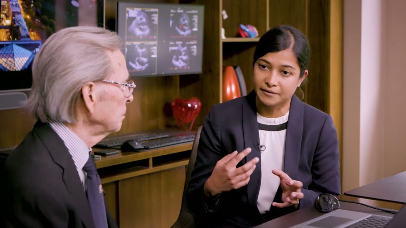
Reviewing surgical video from a case study, we explore the complexity of diagnosing mitral valve endocarditis, especially when there is a risk of embolism. In this discussion we review:
The key role that echocardiography plays in accurate diagnosis and management of native mitral valve endocarditis
The importance of thorough CT exams to evaluate the possibility of systemic and cerebral septic embolic events
Optimal timing and technique for surgical intervention when there is a risk of cerebral embolic events
To refer a patient, please call the cardiovascular surgery referral line at 212-659-6800, thank you.
Hi, I'm Doctor Randy Martin. And we're here at Mount Sinai in New York. I'm a clinical professor of cardiovascular surgery here and I'm thrilled to be joined by Doctor Artie Patel. Artie is a cardiac surgeon and also assistant professor of cardiac surgery. And Artie, uh, endocarditis is something I've been interested in my whole life as an echocardiographer. But you've got a really interesting case. Tell me a little bit about it. We do. Yes. We actually had a patient who came in with native MIB endocarditis a few months ago, uh, for which we performed a MIB repair and that's something I want to talk a little bit more about. So this was a 51 year old, otherwise very healthy gentleman who had some dental work done, uh, probably about two months ago before he presented and in two weeks before he came to us, he started having some very low grade symptoms, some fatigue malaise wasn't really able to get through his whole day without needing a lot of laps, naps throughout the middle. And towards the end of the two week period, then started having low grade fevers, which got his wife very concerned and she brought him to the, er, in the, er, they just did a full set of work up X ray EK GS labs and found his, in fact, his white count was quite elevated and just his routine ended up sending blood cultures which a few days later come back positive. So he was admitted he was with the concept of, he had endocarditis. Well, actually it was just back to Ria and then one of the, one of the hospitalists thankfully listened to him and heard a murmur, uh which led to a little bit more further work up. They got an echocardiogram that showed um mitra endocarditis with severe micro regurgitation. Ok. Ok. And then what happened to him because the story gets unfortunately more complicated. It does. So we were consulted at this point and this was at an outside hospital and he was being transferred to us to get for potential MIB repair given that he's a very young, healthy 51 year old gentleman. And the day he's about to be transferred over starts having very severe headaches. So everybody's like, ok, let's wait, let's wait to see what's happening. Let's work this up a little bit more. Um They were concerned about a stroke given that he had endocarditis and pretty decent size vegetation in the valve which we'll look at in a minute. Um His work up, we got the CT head and the CT angiogram actually showed a mycotic aneurysm and also a small subarachnoid bleed. So he had one of the dreaded complications of endocarditis, especially with, with big left sided valve vegetations and an em, embolic of it to his brain. Ok. So, so, so now he's got this, he's got the subarachnoid hemorrhage. People always used to worry a long time about doing surgery on them because you put them on the, on the heart lung machine and they could have excessive bleeding. But you all, what was your thoughts here? You, I see that you waited to have him do an antibiotic, a complete antibiotic regime, but you didn't wait long. He waited about three weeks, three weeks. Yeah. So he actually did, ended up getting uh a coiling for the mycotic aneurysm. But given the fact that he'd had a subarachnoid hemorrhage and the fact that the patient was actually extremely stable, completely asymptomatic was able to do his day to day stuff, walk around, go up and down a flight of stairs without any shortness of breath. We elected to actually wait a few weeks just to be far enough away from the neurological event to reduce the risk of any bleeding from going on bypass. Since we do have to apron on bypass, going to show me his uh echo here. Absolutely. So here's the echocardiogram and pretty severe mi regurgitation, posterior leaflet flail. You also see a pretty big vegetation that's flopping in and out of view, quite mobile. Actually, the interesting thing here is that you, yeah, he does have a big veg, but he's also got a big left atrium which tells me that he's had macho gur for some time. So, and he's got, you know, you can see uh uh a jet that's really a complex jet, but it's directed anteriorly, but then uh in another direction too. So this is this veg is on the posterior away one and it's big. So you all see this, what, what are your thoughts now? So again, one of the other reasoning was OK. This is not a cue to my regurgitation, just like you pointed out, the, the ventricle is quite big and large. He's probably had had it for a little bit more longer than he realized. That was one of the reasons we decided to wait, give it a little time, make sure the infection is cleared out if possible before the surgery, that way any prosthetic material we use has less chance of getting infected as well. So then given that he is a young, healthy person, any kind of as long as the leaflets have good mobility. And there is enough leaflet tissue. I think we always like to consider repair, especially in younger patients in the hopes that this is hopefully the only surgery they will need throughout the course of their lifetime. So if patients in my experience with patients who get systemic emboli, they can get emboli to other organs, display your kidney other things. Did you all look for that? Yeah. But actually as routine, any time we have anybody who comes in with endocarditis, we like to get a head ctct of the chest abdomen for the exact same reasons. Very often we'll see embolization to the spleen or even micro embolization to the brain a lot of times on every day. And that helps you with your management of knowing what to do with these patients. So, so with this gentleman, you did surgery on him. We did, yeah, we did do surgery on him and bringing the point about embolization, we've actually had quite a few people who initially get, we get consulted on being like, ok, endocarditis, maybe they don't need surgery right now. It's a large vegetation but only have moderate micro regurgitation. But you then go ahead and do the imaging and find evidence of embolization, which changes the indication the surgery because if they just had a moderate leak, you probably would have just waited it out. We, you know, we in the early in the early days of echo when we started seeing these big vegs and we were, we were very concerned about, about embolization and it was really hard to get others. There's this concern until until really the awareness of the extent of systemic embolization these patients and the consequences of that. So I think, I think being able to know not only where you are but the extent of the e especially cerebral imp conversation and knowing the best time to operate on him is really important. You have some uh interesting pictures from the ori I actually have a nice little video. So here this is uh the big vegetation that you see on the posterior leaflet here all the way on the under surface of it and a completely ruptured cord. Uh It's a big flail P two segment here. This is just the jet lesion from the regurgitation again, the fact that he's probably had the regurgitation for a little while longer than we realized. So you're going to do because this looks localized. Are you going to remove? Yeah, we're going to try to repair. I mean, if you look at the rest of his leaflets, actually, they're actually very pliable, not completely destroyed. So definitely it's something repair is something we would consider it. Ok. So here you can just see that the rest of the P two segment is quite diseased, but the P one and P three segments actually look quite good. They have good healthy cordia, good healthy tissue. So here we are resecting the infected P two segment just again, making sure we have a wide resection, we don't leave any infection behind. Um And then we go ahead and like put the leaflet back together. So you're just gonna do a, a primary repair just as you would with just a bit. Yeah. But, but the, but the, the point is, is that you knew that this was localized and you could remove that. You didn't see any signs of any other involvement at that point in time. So you knew that the repair would be the right thing to do for him that, you know your thoughts about doing a mitral valve replacement versus repair. Absolutely. I think it's always reasonable to consider a repair. It's not uncommon sometimes. Ok, we'll think about, you go to the operating room thinking, ok, we're going to go ahead and do a repair, but you go in and find extensive tissue destruction, then you don't really want it. Again. That's a debate in terms of how old is the patient. If it's a younger patient, you can reasonably do a good repair that you're confident is going to stay, keep them out of the operating room and needing another surgery in the long term, then it's definitely something you can attend. But if it's an older patient or like a sicker patient or if there is extensive tissue destruction, you don't think it's going to be a durable repair, then I think you have to weigh the risk versus benefit. And in that case, a patient might be better off with a replacement and you, you know, you're going to weigh the having uh more artificial material in the heart in those patients. So, having them is well treated with antibiotics in both of these cases, but is really important, but especially that. So you've used a, a band in here and you? Yeah. So we, this, we used a band, we did the P two resection, um close that segment up. Yeah, I also had a little bit of a cleft which we closed just because of the residual leak. Um We use a neo cord to kind of fix the height and then the band to kind of stabilize the repair. And you see the echocardiogram here actually shows pretty good uh micele. So what are your, what are your take away? Did the gentleman do? Well, he did great, did very well routine post operative course, went home after five days. Um, a lot of time with endocarditis, especially if they've been recently treated. We do elect to do another few weeks of IV antibiotics post operative, which we did just in case of any residual infection or anything we might have stirred up in the operating room from Debre the infected areas. Good. So what are your take home? So, the big thing I think is uh microvalve repair should always be considered in patients with endocarditis. Um As long as you don't have an extensive tissue deficit, I think it's a reasonable consider. It's a reasonable consideration for most patients and especially younger patients who have 1020 3040 years to live. Um It's worth giving them a repair that will hopefully prevent them from having or will help them to not need another surgery further down the road and every time you do a mi endocarditis repair, you have to be sure you have good source control in the sense that all the infected tissue is removed. You leave some infected tissue behind. It's going to come back to halt you. Very. Yes. Either it's not going to the repair is not going to hold or it's going to get reinfected. So you did, I'm looking at your second bullet point there. So you did, obviously you take out the infected tissues targeted, but tension free leaflet reconstruction. So you're doing the, you're do, you're using the same techniques that you use in a, in a non infected mitral valve. You really wanna have good coaptation, deep coaptation and no tension on any, any structures. And, and it's, you can do that in there in repair. You're saying lower is the reinfection rate you're talking about versus more uh artificial equipment in there like a total mitral valve replacement just because you have a lot more prosthetic material um in a replacement versus you have in a repair and having your own tissues in terms of the valve leaflets, I think makes a difference too. Yeah. You know, I, I keep referring because I, I fought this battle with uh ID for so many years. This concept of early intervention in big mitral valve edges that, that have a lot of Mr and, and mentioned back in, in early 2009 or 2010, Jim Gamby. Uh and the group in, in, in uh Baltimore published a good series on 90 patients. 89 patients looked at this question of if you have cerebral embolic events, which they did, they had a fair number in some subarachnoid bleeds and found that if and they, their main period to operate on these patients was four days. So they did really early surgery, they might have been much sicker than your patient. But they said if you only had less than a centimeter or two of, of surrounding hemorrhage, that your chances of bleeding, because that was really the other risk. If you put them on the heart lung machine, are they gonna bleed? And so their outcomes is really good. Their five year survival was like 90%. So it's really good. So exactly what you all did, you had time, he was stable. Um, you know, for people who are watching this and thinking, uh if you have a patient who's, who has uh valve endocarditis, especially mitral valve endocarditis, what do you need to think about? I think the big thing is always think about one is embolism always rule out that there is no embolism to the brain, to the spleen liver, kidneys very frequently. I have seen consoles that we've gotten in patient is we do, the patient looks completely fine. There is really low for surgery, looking at the echocardiogram. Ok? The vegetation is kind of small but you might have cut the vegetation later on. But you do a pan scan, scan their head and there's already micro emboli, there's already some splenic inox renal inox. And that has changed management. I think it serves a patient better in these cases to be operated upon earlier. Yeah. And, and your point is get, get the infected tissue out there. And it's, it really is, it really is important, I think to do exactly what you said to look for for em embolic things because it changes your management. But this is a great case, tremendous illustration and most important the young man is doing well. He's doing fantastic. His post operative follow up has been great. Thank you. Thank you very much for um you know, telling us all about this. I hope you enjoyed this case. A lot of great teaching points and, and good clinical pearls. So thanks for joining us. Thank you.


