Mitral Annular Calcification (MAC) is a complex and increasingly common syndrome. In this discussion we review Mount Sinai’s proven algorithm for determining the best treatment for MAC, as well as imaging and intraoperative videos that highlight:
The incidence and etiology of MAC, especially for those at high risk: the elderly, patients with CKD, women, and patients with Barlow’s mitral valve disease
The critical role of CT imaging to quantify the extent of mitral annular and valvular calcification, and its location in the annulus, leaflets or ventricles
Key surgical considerations, including:
Periannular suture technique
Management of the anterior mitral valve leaflet to reduce the risk of left ventricular outflow tract obstruction
Proper selection of mitral valve prostheses
Why referring patients with complex mitral disease to designated mitral valve reference centers improves clinical outcomes
To refer a patient, please call the cardiovascular surgery referral line at 212-659-6800, thank you.
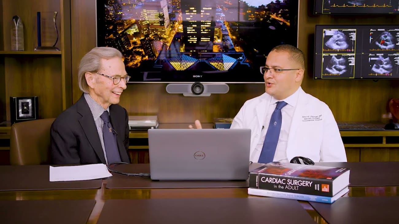
Hi, I'm doctor Randy Martin. We're here at Mount Sinai and I'm a clinical professor of cardiovascular surgery. I'm thrilled to be joined by a colleague and a dear friend. Ahmed. L is Maui Ahmed is the clinical director of the Mitral Reference Center Reference Repair Center. And I think that's fabulous. You're also associate professor of cardiovascular surgery and you're a fabulous surgeon. I've already said that I can, I can attest to that, but you're gonna, we're gonna talk about a condition that we're seeing more and more of which is Mitra annular calcification. Um, tell me a little bit about that because it is, we see a lot of it. It's an increasing problem. Well, uh, thanks for having me, uh, today, uh, Randy, that's an honor for me. And, um, it's a privilege also to talk about this important topic now, in the field of cardiovascular medicine and surgery, which is Mitra Annual Calcification. Um, it has been sort of a hot topic. Recently. We have seen a lot of presentation on that in almost all the forums in both surgery and cardiology and there are several reasons for it, which we will go through as we talk, but it's definitely a common clinical encounter that we see in our clinics almost on a weekly basis, especially in big centers like Masai. And it's not new from what you know, this is an interesting study as I was looking into the literature just to see when this all started. And then I found the oldest patient who had mac, this patient was 3000 years old. So this is technically called the horror study where they studied almost 52 Egyptian mummies, they did cat scan and they looked at cardiovascular risk factors. And interestingly, two of these mummies had micro calcification, a lot of them had atherosclerosis. Interesting, interesting. So tell us, take us through and I mean, tell us a little bit about what Mac has. So Mac, I would consider it as an age related disease or cardiovascular disease, just like most of the cardiovascular risk factors. It's more common in age population as we know the life expectancy now in developed countries is getting better and better over time, which is something good, but it comes with its own risk including the micro annual calcification, which is technically degenerative or age related changes at the mitral valve level. And the annuals is the supporting structure of that. So you had some risk factors that you've got in there, put people at risk to have, have that and you've got those less than, yeah, there is a lot of cross sectional population study. As we know, beautiful studies done in cardiology and they showed this multi ethnicity acro study, almost every single risk factor for atherosclerosis is assured as a risk factor for mac. But there are several risk factors for this disease also including radiation disease, vision has degenerative might have had disease like Barlow's disease, vision with peripheral vascular disease, smoking, et cetera. So it shares a lot with atherosclerosis and that's what makes it more challenging because those patients tend to get high risk because they have both atherosclerotic disease as well as the annual calcification. But what's more interesting about it as we see more population studies? This is a famous Framingham Heart study and it even showed that the Mac was an independent cardiovascular risk factor for mortality. So, and it's very interesting because you can see the linear relationship between Mac and the cardiovascular risk even for each one millimeter increase in the. So that make the cardiovascular risk increase by tenfold. What makes it, this is very serious? So, it's serious and what it does to the mitral orifice and all that. But it's also a marker of, of increased mortality. Absolutely. Yes. Ok. That is interesting. All right. And is it more common in women than men or it is? Yeah. As we know, and we learned that as you know, the population is aging, we see it more frequently in females. Why are you telling me here? So this is, I'll take it back to surgery because we showed it how the cardiology community, I mean, you're a cardiologist. So I'm sure you are very familiar with the previous studies. But we as a surgeon, we look at it from the technical standpoint because we know we know the patients are sick. But technically speaking, it is a very difficult problem to deal with. Historically, the mec has got a very bad name in cardiac surgery. Unfortunately, the earliest description of major disasters that happened in my replacement was described in patient with Mac. And that's technically what we describe as an ev groove disruption or micro annular, you know, disruption or groove dissociation, different types and different studies describe that. And unfortunately, all these patients started by having Mac. Uh and that's the first thing that we should keep in mind when we operate in this patient is basically how to prevent surgical complications. Ok. So it's common, I mean, it's increasing and there are risk factors for it, but it also is a difficult problem to tackle. Ok, if you're gonna, if you're in there doing an operative repair or doing something on the valve. So keep me moving through this and obviously what, what else you're gonna show me? So, yeah. Yes, I I mean, I collected a few slides today, but I also want to make sure that now it's an epidemic almost. If you do valve surgery in big centers and mi reference center, you see a lot of patients get referred from everywhere. Because the general cardiac surgery community, they try to send those patients to big centers where they have a lot of experience with these difficult cases. So now we have this epidemic of patients coming to us because they have micro valve disease and underlying mac and we have to come up with a safe way or a sort of a strategy or an algorithm about to handle how to handle these patients safely because it's not easy. And there is no consensus about it among us in general. You and many of us know what a normal mitral valve. The top picture you're looking at, you're working from the H right on to the mitral valve. But in this, this patient all all around the valve, maybe almost 360 degrees is calcification. Is that right? Exactly. So, uh yeah, I picked up one of the most impressive cases that we have done here at mons patients had complete circumferential calcification. So you can see the mitral valve is ossified almost. Yeah. And it's pointing out another problem that we're seeing with this is that. So it's really calcium grows in on the aneurysm and then gets on the wale and can narrow the, the orifice so that they become stenotic. It's almost like exactly rheumatic module. Well, disease and few of the mac patients, I think in most of the recent show about maybe less than 10% of patients whose mag they will progress to this so severe miter stenosis. And I found those are the highest risk patients among patients whose mac it's like you're going out and trying to operate on the middle of the street with concrete there. So these are the risk factors. So show me what else we have learned about this. So this is, as I should say, it's a long journey. It started almost 10 years ago here at Mount Sinai when we started to tackle that difficult problem. And we have learned many lessons, I should say the very hard way, you know, just going back into the early cases. The first lesson that we learned is imaging is key. Now the cat scan is mandatory and there are several studies on that. We prescribed our own classification of Mac. As you can tell, it's not only the circumferential extension of Mac, but also the vertical extension if it goes down into the virt as well. And that's what we will correlate primarily with surgical complications and risks. And the important thing you mentioned is is and I should have said that at the outset that you all have had a 10 year experience with this. And the thing when I was talking to you earlier, we're talking about a condition, but you're also always focusing on what's best for the patient. What's then you've learned a lot of lessons in dealing with how to treat this. Exactly. So I mean, as you mentioned earlier, we have to look at the anatomy part of it, which is basically what we're going to do for the matter of that. But more important, I have to look at the patient because those patients are high risk patients, a lot of them, they have really short life expectancy and high risk for bi operative complications. And, and as I'm going to go through the survival et cetera later, we will see that we have proved that those patients have more long term and medium midterm outcomes. So you're working and when you're doing the cat scan, and I think that's really important. You're working at not circumferential and the extent and the, but you also get some idea again, going back to this concept that if it keeps growing in, in the South where I'm from, Katsu is a vine that grows in. But if it keeps growing in, it causes narrowing of the orifice, you could actually measure the how narrow that orifice is. So you're getting a lot of information. Yeah. Yeah, we do a lot of work on the cat scan reconstructed. We have even virtual valve as, as you in the top right corner, we screen those patients in several, you know, transcatheter techniques and, and procedures before we jump into surgery. Of course. So let's go ahead. So again, we were finally came up with an algorithm about to deal with Mac and as you see, it's a little bit of a complex diagram, but we always started the operative candies is a patient operable or not. And there are several things that we look into this, including frailty index comorbidities, even eyeball test, which is something very subjective, but we trust it sometimes. And then if the patient is a potential surgical candidate, then we look at the anatomy assessment which is primarily dependent on the cat scan. And based on that, we can come up with a plan whether the patient can be dealt with percutaneously in the Cath lab or as most of the patient, unfortunately, eventually come to us because as we know, the technology did not really evolve quickly in that field because of the complexity of the micro lab. So you and I understand that the transcatheter approach would be doing this in the Cath lab with a transcatheter deployment in that in that area. And there's been a lot of uh Doctor Guerra has done a lot of a big study on that and the results of that have shown that it's really high, high risk that that tells me the outcome of that is not good. It has not been good. Yes, it is because as I said, it is a high risk patient profile. And there is a lot of technical issues also that you can't really overcome on catheter based versus in open surgical setting where we can have more versatility techniques. So if you go through this and you find out that if, basically, if they're inoperable, then uh then you're not gonna, and there's certain people that you would not do anything for. Is that correct? Ok. And who, who would you do? You've got other choices in here. You've got hybrid, hybrid, uh that's, that's doing a putting in a trans catheter valve. But during an open operation. Yes. Correct. Yeah, that's also an option. So if you come up to the surgical algorithm, this is a much symbol of diagram to understand. So we do the cat scan and then we decide about the degree of the calcification or the class of the calcification. If you have anything circumferential or what we call horseshoe calcification, those patients become more amenable for the open use of the hybrid devices like a verve. For example, it's an off label use of the device, but we reported one of the earliest cases in this field. And now it's sort of seem to be a good option for this patient, but under very selected as it's selected. And you've got, you're going to show me in a minute the technique that you do because what you're trying to do is to see this and a calcified a very hard area. So there's no weak per your wake is not a good jacket on the left. You said that you've also developed when you would do a standard mitral valve replacement. So that means doing something with the calcium that basically has been always seen as a standard of care as we know from the early reports. And cardiac surgery, surgeons used to be very brave and decalcified the mi and anus and put a new valve in and that will give you the best chance for healing in long term even outcomes. But now we have to be careful because remember the patients we see in those days are different from these historic Russ of annual decal. We see a lot of high risk super high risk patients, octogenarians, patients on dialysis renal disease, co morbidities, etc. So we tend to leave the calcium and work more around the calcium rather than decalcified the annulus. And that's to limit the surgical mortality and morbidity. Let's go forward. You keep in mind this is like very few again videos that show how we can replace all good sutures. So tell me in the audience go, can you go back for a second and do that? I know it's quick and keep them short but that it's a short video. But we try to leave the calcium bar and go around the calcium with sutures using big needles. So that's called the very annual suture technique. We use it occasionally and this is just a standard valve and you can see the echo perfect result, no butter valve. And this is a standard valve you put in it and suture it around the calcium. You didn't do anything to the calcium. No we left it alone. The second technique that we, this is again another surgical strategy that we adopted here at Mount. So you can see these valves are calcified, degenerative valve. We tend to replace those valves rather than repairing them because we know the poor durability of repair. And in this case, we call the interior leaflet flip. So we took out the anterior leaflet and flipped it to the to create sort of a new analys. And again, we go for a standard valve and we have a good result. So that's our to go technique or our preferred option is to use standard surgery as much as possible. And basically, you're putting on a standard artificial mitral valve there. That is the TSS prosthesis that you put in there. But using standard surgical techniques. Absolutely. And we have a lot of experience with this. Now, as we are doing a lot of these cases because we know that if you do that, we have a sort of more predictable outcomes and durability versus as opposed to the new techniques which we, we will talk about. Ok. So this is again, you were talking about the te valves. So this is a patient with a circumferential mac. Again, there is a lot of technical tips here, but we have a lot of things we can do which we cannot do in the Cath lab. We took out the interior leaflet, we can put some sutures to prevent imb we can wrap this valve in teflon filth to prevent parallel regurgitation. I mean, you can't do any of this in the Cath lab and that's what makes it more challenging. And again, we have a good result in this patient, but I should say this is off label use. We don't know, we don't know the durability. It definitely improved the quality of life in the patient and it has to be done very selectively. So this patient had severe and it worked like circumferential back and then it was all, it had mitral stenosis in essence. So you took out, you did two things there, I think is what we wired from the transcatheter is that per A are really bad. And you could sometimes you could get a equation of by the anterior A foot would include the LV outflow track when you shove it up there. And that's those cause more t those are the achilles of this technique because there is not as you know, a simple way to fit to, to deal with this uh complications. You took out the anterior away foot and then you're making these to all the way around it. So you are trying to see it in there the best possible way because you know, we don't use any switch. This is a balloon expandable device. We basically trust the radial tension of that valve to stabilize the device in place just similar to the field. But this is completely different from the valve because this valve is exposed to systolic pressure. So it's not like an aortic valve with it closed on the dito pressure. So a lot of physiological and anatomical variations that make the mitral valve is a much more challenging to deal with. So who gets the hybrid? In other words, those, when would you determine? So you had, you given me two choices, you could do a standard uh replacement and then you could do a tower and open open surgery, but you're going to do a tow tower type valve who gets the tower type be to valve is always a bailout. So what we do, we do the measurements, we know it's anatomically feasible, like either the annulus, all the measurements are good. Um As you know, Gilbert, I mean, he's so we, we do these cases in collaboration. It's as you know, it's not only me, we have to clear these patients before we go to the or we go as well as a stand by. And we, we look first, we do our surgical valve analysis. We will always go for a surgical replacement if we can do it because that's the more predictable and safer way to handle these cases. I'm showing you the best case I've done, but I didn't go through the learning curve where we have seen every single communication possible. So you told me in a conversation we had a couple of months ago that this early on. I mean, this is really heroic surgery. And he, I mean, patients are really quite, but it was the sense that, that you were trying something and your, your results were not good and you really wondered whether you should even be doing this. Exactly. At some point, you know, like any novel procedure you try, especially in high risk patients, you, you have to face a learning curve. There is no, you can't keep it unfortunately. But the key thing is you were thinking about the patient there. I mean, you were thinking about the outcome of the patient, not just doing another procedure exactly early on, we were sort of geared towards more technical success. But then we learned across the years that it is not only the technical deployment of the world, but also the long term outcomes of the patient and the improvement of quality of life and avoiding the communication as you mentioned. And two things, one is that you, we have wired that I I sent you a paper that just was out of the echo and the cardiology is that this like the patients you showed there has got extensive mac that's on the annul its on the, they get basically stenosis, they get narrowing of the or those people have a horrible outcome. I mean, a f 50% mortality in two years or so. And that's, that's a really, that's a tough one where this is this is a procedure you would do the, the question, I'd have not everybody. I mean, you're, you're showing us people with extreme complex Mac. What about somebody who has mild or moderate Mac? You're doing a mitral valve repair? What do you do with that? Mild or moderate Mac? There is many things you can do. Mostly we take, we take the Mac out to improve the durability of the repair because, you know, we repeat a lot of Monza so mild to moderate Mac, it shouldn't be an issue. You take it out are you, we would rather, we would rather take it out if it's multiple, especially in young patients because that will be translated to more durability and more healing around the. And that's the kind of ma that doesn't extend down into the V and all the ones that put the patient at risk. So that's why I think CT scan almost begin the standard of care now. And so you've done this, um as you said, you've had 10 year experience, you've got very large series. But one of one of the lessons that, that you've learned or what would you like the viewers of their cardiologist or cardiac surgeons or even consumers to know uh what, what lessons have you learned, who learned a lot of lessons? I mean, this is again, this is a patient. I just showed the video. Um this is one of the patients who came to us from Florida she was 84 but in good shape, she passed the eyeball test, we did the cat scan, we saw the measurements, we thought that she would be a perfect and she had a great outcome and she really had that transformed her quality of life. So I guess that's Gilbert, that's Gilbert. I don't want to forget Gilbert. So, so Gilbert has been keen the success of that program here in Mac. He's a colleague who's a surgeon but trained in, he's, I don't know how to call it, but he, he call himself structural surgeon or surgeon interventionist. I asked you about the last word, what I mean? You know, what, what, what, what do you want people to know about this? Well, first, as I said, it's not, you have to be careful because these cases, I guess knowing them in, in, in the center is important because we develop um uh experience and, and, and I showed that here in the double ET meeting a few weeks ago, you see clustering of communication early on during our experience, we started almost 2013 just starting doing this. Um And then before we have a sort of a cluster of, of, of, of straightforward cases and then now you still can't see some communication, but you would be good in learning how to deal with complications and how to get more complications or. So there's a learning curve and people need to either, you know, reach out to experts in the field or see experts in the field if this is going to be done a other lessons or is that, that sounds like a pretty good lesson? Yeah, we also look at the long term outcomes. I mean, this is a one year outcome that showed even patients who survive the one year survival is not necessarily uh perfect on these patients, particularly if you get A T the one year was almost 50% versus almost above 80%. The B had a standard valve. I think the explanation of that is that those or how much calcium we have because we know that we do the TB has more calcium and we know from the Framingham started that are higher cardiovascular mortality and, and poor survival. Uh So this is an again uh uh listen to learn, I mean, you really do what you, what you've told me is that you, you know, like everything, if you're a physician who's interested in not only helping the patient but learning and improving the technique is that you aren't over time, but that the imaging really is important to tell you the extent of the calcification, its location and depth and then you then you go through certain choices that you would have. Is this something that where people should refer patients for who have extensive calcification to reference centers? I think it's mandatory because we have all the infrastructure for imaging and valve analysis and hyper techniques and transcatheter options and also clinical trials. If the trial, there are several trials running for MAC now. So we screen those patients and we do the homework before we come up to the decision, whether to operate or not. This is, this is really good. I mean, I appreciate you sharing this with me. I know that we've been seeing it on echo for years but but but really imaging but mainly CT scanning is really the key. Now, 100% telling you how to, how to advance the patient. Definitely, I think it's a key of this field is imaging. A lot of this conversation. As I remember, we didn't have good imaging at that time. And that's why we didn't have sort of predictability of complication, including outflow obstruction, leak, circumference of the valve, etcetera. So everything comes together, I think as the field moves imaging, cardiovascular interventions and surgery, we we become better eventually. And it's really, really important. I mean, if you've in being here at the, at the Metro Reference Repair Center, that that's really what you're doing and you've learned a lot. So thanks for sharing. You know, I always learn a lot that the surgery. This is uh yeah, we will end with this slide. I mean, this is a good article that we wrote about surgical management to make it basically summarize our philosophy or concept and, and different techniques. And now you can now you can extend it as you've got. That's, that was in 2020. So now you've got more patience and some extension. Exactly. So we probably can rewrite the question. Thank you. Thank you very much and thank you for joining us. I hope you've worked as much as I have. Thank you very much, Rick the.


